ICAT 3D Cone Beam CT
- Home
- ICAT 3D Cone Beam CT

ICAT 3D Cone Beam CT
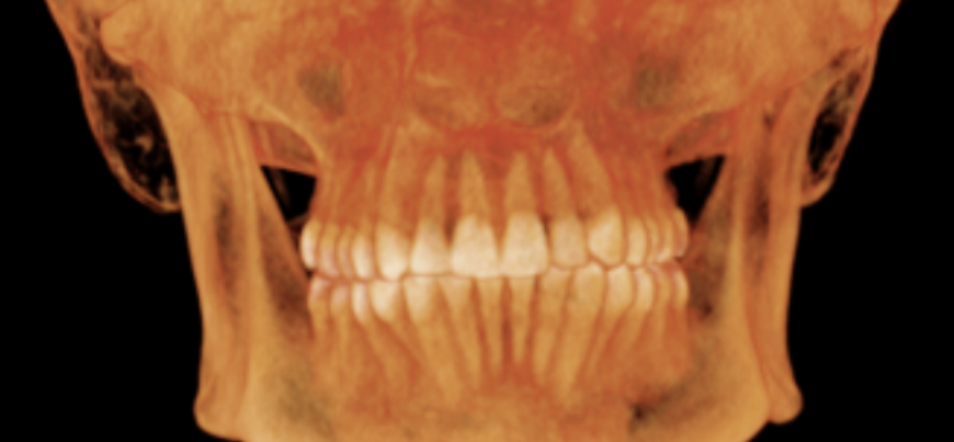
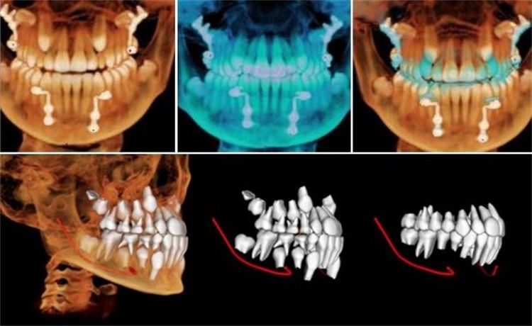
In a Cone Beam CT (CBCT) scan, a cone-shaped X-ray beam is rotated around the head of the patient, exposing a sensor to multiple views of the teeth and jaws. A computer then renders those scans into a volume of images of the region of interest that are then reconstructed to allow viewing slices at any angle or thickness.
More importantly, CBCT allows viewing teeth in ways that cannot be seen in periapical radiology. CBCT also displays bone and bone defects. This new level of detail allows for more precise treatment planning.
It differs from spiral (fan) beamed medical CT in that it is lower in cost, and it exposes patients to less radiation.


